Monday, 19 October 2015

Salmonella spp. from Blood culture

Salmonella are predominantly motile (most species except Salmonella pullorum-gallinarum are motile with peritrichous flagella), non-spore forming, Gram negative rod bacteria that belongs to Enterobacteriaceae family. They are divided into two species – Salmonella bongori (previously serotype V) and Salmonella enterica (Salmonella enterica is subdivided into subspecies – enteric (serotype I), salamae (serotype II), arizonae (serotype IIIa), diarizonae (serotype IIIb), houtenae (serotype IV) and indica (serotype VI),  and over 2500 serovars. The serovars are defined based on the somatic O (Lipopolysaccharide) and flagellar H antigens (The Kauffman-White classification).
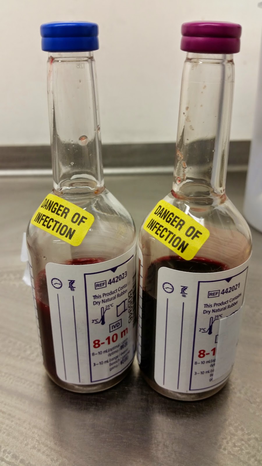
The blood culture aerobic and anaerobic bottle flagged up positive after 8 hours incubation in the BD Bactec FX Blood culture analyser and upon Gram stain shows Gram negative rods. Routine blood culture plates where inoculated according the Laboratory SOP - Blood agar, chocolate agar, UriSelect chromogenic agar and FAA AN anaerobic plates. All culture plates were purchased from E & O Labs (http://www.eolabs.com/). After 18-48 hours incubation at appropriate temperatures and condition, there was growth on all plates. The Salmonella isolate on culture plates are shown below.

Culture medium
Incubation condition
Incubation Time
Appearance
Blood agar
CO2 at 37OC
18 – 48 hours
moist, grey, round non-haemolytic colony
Chocolate agar
CO2 at 37OC
18 – 48 hours
moist, grey, round colony
UriSelect chromogenic agar
Air at 37OC
18 – 24 hours
moist, white/cream, round colony

This isolate was then identified using Biomerieux Vitek MS (MALDI-TOF) as Salmonella group. Purity plates were done on CLED plates and used for agglutination test after 24 hours incubation at the appropriate temperature condition. Salmonella isolates are used for serological confirmation using the agglutination test Polyvalent O and Polyvalent H specific antisera. This is an easy, quick and straightforward method. Specialist reference microbiology laboratories use more advanced and automated methods such as PCR technique.
The Kaufmann-White scheme is a system complex serotyping scheme that classifies Salmonella into serotypes based on their surface antigens. Specific antisera are raised against three antigenic sources; the ‘O’ polysaccharide component of the bacterial cell wall, the ‘H’ flagellar antigens present in most strains and the ‘Vi’ virulence/capsular antigen present mainly, but not exclusively, in S.typhi. It was further found that each strain could possess several ‘O’ determinants and so could be attributed to a particular ‘O’ group. Additional subdivision could also be made on the basis of flagella ‘H’ antigens as Salmonella exhibit phase variation between motile (phase 1) and non-motile (phase 2) phenotypes. The Kaufmann-White scheme was first published in 1934 listing 44 serotypes, but now contains serotypes for more than 2500 Salmonella spp and is used both in routine and specialised laboratories for the identification of Salmonella species. Examples of the serotypes can be seen on the table below


Serotypes

‘O’ Antigens
‘H’ Antigens
Phase 1
Phase 2
Salmonella typhi
9,12,Vi
d
None
Salmonella paratyphi A
1,2,12
a
None
Salmonella paratyphi A var. Durazzo
2,12
a
None
Salmonella paratyphi B
1,4,5,12
b
1,2
Salmonella paratyphi B var. Odense
1,4,12
b
1,2
Salmonella enteritidis
1,9,12
g,m
None
Salmonella typhimurium
1,4,5,12
i
1,2


Salmonella spp. on blood agar
Salmonella spp. Gram negative rods
Salmonella spp. on Chocolate agar
Salmonella spp. on UriSelect chromogenic agar

Wednesday, 26 August 2015

A brief description of Eubacterium limosum from Blood culture


Eubacterium limosum
Eubacterium limosum is a Gram positive, non-branching and non-spore forming rod that grows in anaerobic environment. Eubacterium spp are commonly seen as a normal flora in the gastrointestinal tract of humans but can cause bacteraemia in rare occasion.
This organism was isolated from the blood culture specimen of a hospital in-patient with clinical details of diarrhoea. The BD Bactec FX Blood culture analyser flagged the anaerobic bottle (pink top) positive after 56 hours incubation and upon Gram stain shows Gram positive non-branching rods. The specimen was then plated out on Blood agar, chocolate agar, UriSelect chromogenic agar and FAA AN anaerobic plates. All culture plates were purchased from E & O Labs (http://www.eolabs.com/). After 24 hours incubation at appropriate temperatures, there was growth on the FAA AN anaerobic plate only. The isolate shows a smooth, rounded, lustrous colony as seen on the culture plate above. This isolate was then identified using Biomerieux Vitek MS as Eubacterium limosum.

Thursday, 19 March 2015

Urinary tract infections (UTI) 2

Principles of three different automated urine microscopy analysers - Sysmex UF1000i, Menarini sediMAX and Iris iQ200 
The use of automated system in urine analysis reduces the time and cost used in the manual routine analysis. In addition, automated system analyses a high number of samples in a short time (up to 100 samples per hour). There are a few methods available for automated system which includes the Sysmex UF1000i, Menarini sediMAX and Iris iQ200.

The Sysmex UF1000i method uses the urine flow cytometry technology to detect red blood cells, white blood cells, bacteria, epithelial cells and casts in urine. The automated system dilutes 200µL of the urine sample with citrate buffer solution and cationic surfactant with an addition of 40µL of fluorescent dye to complete the mixture. This mixture is then passed through a sheath of flow which is exposed to a laser beam to detect the particles in the stained urine sample with the fluorescent dye staining the nucleic acid in bacteria. The identification and characterisation of particles are based on the fluorescence intensity, forward scatter light and side scatter light intensity which generates electrical signal patterns by photomultiplier conversion which is then measured.

The Menarini sediMAX is an automated microscopy image based sediment analyser used in urine microscopy. The analyser produces a monolayer of urine sediment by centrifugation in a special cuvette. The sediment is analyzed by a bright field microscope and digital camera to capture and categorize 15 particle images based upon size and shape using image processing software. The digital images can be viewed by a trained laboratory staff with zoom capability. The instrument has a high sensitivity (>80%) in the detection of RBC and WBC but has a low sensitivity (approximately 50%) when compared with manual microscopy.

The Iris iQ200 method uses digital imaging and auto particle recognition to classify and quantify urine particles in uncentrifuged urine based upon size and shape. It classifies images such as WBC, RBC, bacteria, casts, yeast and crystals into categories. The digital images generated are collected, approximately 500 frames per sample, then viewed and classified by a trained laboratory staff. This is usually achieved without the use of manual microscopy.
The advantages and disadvantages of the automated urine systems (Sysmex UF1000i, SediMAX and Iris iQ200) are listed below.

Analyser
Advantages
Disadvantages
Sysmex UF1000i
Uses uncentrifuged urine samples.
Improved bacteria counting and flags for UTI.
Urgent urine samples can be processed by operating the analyser manually.
Throughput of 100 samples per hour.
Cannot differentiate clinically significant pathologic casts cell from benign casts.
Manual microscopy is carried out on flagged results.
sediMAX
This analyser does not require any reagents.
Requires minimal laboratory technician training to interpret images that are similar to manual microscopy.
Results may be available for viewing from the stored digital images
Throughput of 80 samples per hour.

Yeast is mistaken for RBCs.
Epithelial cells are mistaken for WBCs.
Particle loss may occur due to the ultracentrifugation of urine samples.
Iris iQ200
Uses uncentrifuged urine samples.
Accurately quantifies bacteria.
Results may be available for viewing from the stored digital images
Throughput of up to 101 samples per hour.
Crystals are mistaken for RBCs.
There is no sufficient resolution in order to classify all dysmorphic RBC variants which prompt manual microscopy.
Requires considerable laboratory technician training for viewing digital images that are different from manual microscopy.

Detection of biochemical markers and their significance in urine samples
Biochemical markers in urine such as glucose, bilirubin, nitrite, ketone, specific gravity, blood, pH, protein, leukocytes and urobilinogen can be used for UTI. It can be done conveniently as a point of care test or near patients test outside the laboratory. This can be done manually (dipstick) or automated (Siemens Clinitek and Roche Urisys 1100).
A urine dipstick Bayer urinalysis strip such as the Multistix 10 SG is a narrow plastic strip with squares of different colours on it which represents each of the biochemical markers present. The strip is usually dipped in the urine sample to be tested and colour changes noted within 1 minute. The results are then interpreted as shown in the table below.

Table 2: Biochemical markers and its clinical relevance
Biochemical marker
Clinical relevance
Nitrite
Bacteria presence in urine
Leucocyte esterase
WBCs presence in urine
Urobilinogen
Possible liver disease
Bilirubin
Possible liver disease or RBCs breakdown
pH
Acidity of the urine sample and useful in interpretation of microscopic crystals
Ketone
Product of fat metabolism
Specific gravity
Concentration of urine
Glucose
Diabetes mellitus
Protein
Glomerular disease
Hemaglobin
RBC trauma or enzyme deficiencies

Automated systems such as Siemens Clinitek analyser is an easy to use chemistry analyser that is used to read Bayer urinalysis strip such as the Multistix 10 SG. The dipped urinalysis strip is inserted into the analyser and result reported automatically.
The Roche Urisys 1100 urine analyser is a semi automated analyser used for in vitro semi quantitative determination of urine analyte. It is a reflectance photometer designed to read and evaluate the results of urine test strips such as Chemstrip 10MD, Chemstrip 7 and Chemstrip 5 OB. The test strip is placed on the sliding tray and a stepping motor moves it under the reading head which remains stationary. The analyser then reads the reference pad followed by each of the pads on the strip. The reading head contains LEDs that emit light at various wavelengths. The LED emits light of a defined wavelength onto the surface of the test pad at an optimum angle. The light hitting the test zone is reflected proportionally to the colour produced on the test pad and is picked up by the detector, a phototransistor positioned directly above the test zone. The phototransistor sends an analog electrical signal to an A/D converter, which changes it to digital form. The microprocessor then converts this digital reading to a relative reflectance value by referring it to a calibration standard. Finally, the system compares the reflectance value with the defined range limits (reflectance values which are programmed into the analyzer for each parameter) and outputs a semi-quantitative result. Each test pad is read photometrically after about 55-65 seconds. In strongly alkaline urine samples, the Urisys 1100 Urine Analyzer automatically corrects the result of the specific gravity test.

Urinary tract infections (UTI) 1

Urinary tract infections can be defined as an infection that affects the upper (ureters and kidneys) and lower (bladder and urethra) urinary tracts. The Health Protection Agency (HPA) stated in 2009 that Urinary tract infections is the second largest healthcare associated infection and estimated that it causes up to 20% of all hospital acquired infections.
Urine microscopy is a method used to identify and quantify cells, bacteria and other materials such as casts in urine samples. This may include the use of an inverted microscope or automated system. Sysmex UF1000i automated analyser is in use for urine microscopy in my laboratory and manual microscopy is rarely performed on urines sample received. This is only done when a request specifically asks for cast cells as the UF1000i cannot differentiate them.  In addition, high risk urine samples are usually processed in Category 3 room without any microscopy performed.
Inverted microscope is a semi quantitative method of urine microscopy. The technique involves the use of micro-titre plate with the lamp or light source and condenser located at the top. The objectives, nose-piece and eye pieces are located below the stage. An aliquot (60 µl) is pipette from a well mixed urine sample and dispensed in the microtitre plate. The plate is then left to settle for 5 minutes and then examined within 15 minutes of dispensing under the inverted microscope using x20 objective lens. The number of RBCs and WBCs per urine sample is counted in two fields and the total number of cells reported as number/mm3. The microscopic results are then reported as <40/ mm3, 40-100/mm3, 100-200/mm3 and >200/mm3. This method is also useful for detecting casts in urine samples. It is also used on samples that cannot be processed using the automated analyser such as samples with thick pus, frank blood and requests for Trichomonas. There could be errors in the use of inverted microscope which includes volume discrepancies and uneven distribution of cells.
Alternatively, a drop of urine is placed on a microscope slide and examined under a normal light microscope using x40 objective lens and the results reported as follows
< 2 cells per hpf        -           < 50/mm3
2 - 5 cells per hpf     -           50 - 100/mm3
5 - 10 cells per hpf   -           100 - 200/mm3
> 10 cells per hpf     -           > 200/mm3

Occasionally, it might be necessary to dilute a urine sample for microscopy if the number of cells present is so large that it masked other cells. Urine samples with numerous white cells are double diluted in saline depending on the number of cells present and then multiply the result by 2. Urine samples with numerous red cells are double diluted in white cell diluting fluid to lyse any red cells present and then multiply the result by 2.

The significance of cells, (other than white cells) casts and pathological crystals that are found in urine are detailed below
Bacteria/Yeast cells 
When there is presence of >1000 bacteria cells per ml in a given urine sample, it usually indicates UTI. This is then followed up by culture. However, differentiation should be made between infection and contamination as normal skin flora could highlight a positive result. These cells if not treated could cause kidney infection by ascending through ureters. Yeast cells are reported as present or not present.
Red blood cells (RBC)
The presence of RBC in a urine sample does not necessarily indicate infection as small amount can be seen in healthy individuals and contamination could occur from menstruation in women and can be visibly detected, by automated urine analyser or with the use of a dipstick. However, in other individuals, high level of RBC in urine known as haematuria may indicate infection especially when the WBC is also elevated. Haematuria can also be seen in renal calculi, malignancy, trauma, stones and glomerulonephritis.
Epithelial cells
The presence of squamous epithelial cells in a urine sample usually indicate contamination from the perianal region.
Cast cells 
Cast cells which are cylindrical proteinaceous structures produced by the kidney and usually present and helpful in the diagnosis of renal infection. They are usually formed in the distal convoluted tubule and collecting ducts of nephrons present in the kidney where they then dislodge and can be seen in the urine. There are acellular and cellular cast cells.
Acelluler cast cells
-       Hyaline casts: Hyaline casts are cylindrical, clear and have a low refractive index. They are produced when Tamm-Horsfall protein aggregates and are usually seen in healthy patients, vigorous exercise or dehydration. 
-       Granular casts: Granular cast occurs as result of either degeneration or breakdown of cellular casts and appears like cigar-shaped, fine or coarse inclusions and with higher refractive index than Hyaline casts. They can be seen in chronic renal disease or after strenuous exercise. 
Cellular cast cells
-    RBC casts: The presence of RBC casts usually indicate glomerular bleeding or damage which can occur in glomerulonephritis. They can also be seen in renal infarction and subacute bacterial endocarditis. They appear as a cylindrical shape with or without ragged edges and yellow-brownish colour.
-   WBC casts: The presence of WBC casts usually indicate kidney infection or inflammation such as in acute pyelonephritis and acute allergic interstitial nephritis.
-      Epithelial cell cast: Epithelial cell casts usually occur when there is damage to the tubular epithelium and can be seen in acute tubular necrosis and when a toxic substance such as mercury is ingested.
Pathological crystal cells
The precipitation of crystals of uric acid, calcium phosphates,Calcium oxalate, triple 
phosphate and amorphous phosphate or urates in urine is called Crystalluria. This 
may be normal, asymptomatic or in association with the formation of urinary tract 
calculi. Crystals may form as a result of drug metabolism and can be seen in 
association with the pathological disease as seen in the presence of uric acid 
crystals in the urine of patient with chronic renal disease and gout. Cysteine crystals 
are rarely seen and may indicate an underlying disease. Bacterial infections caused 
by urea splitting bacteria such as Proteus spp. has been linked with the formation of 
urine stones (Ford 2010).
References
Ford M (2010). Fundamentals of Biomedical Science; Medical Microbiology. Oxford
University Press, London.
Health Protection Agency (2009). Trends in rates of Healthcare Associated Infection 
in England 2004 – 2008. Reports for the National Audit Office. London.